

Excretion and Water Balance
 |
photo by P Matthews |
Objectives
- Gain a knowledge of the mechanisms for maintaining homeostasis in water balance
- Know the structure and function of the malpighian tubules, rectal pads, rectal papillae and cryptonephridial systems.
Topic Outline
- Overview
- Homeostatic Mechanisms
- Malpighian Tubules, Urine production
- Endocrine Regulation, Rectal absorption
Activities
- there are no minilectures at this time
Overview
Maintenance of water balance is a critical issue for insects and other land living arthropods. They have large surface area to volume ratio and are therefore subject to rapid water loss. Protein metabolism produces nitrogenous waste products which must be eliminated incurring water loss with faeces or urine. Insects have evolved behavioural, morphological and physiological mechanisms to maintain a steady state (homeostasis).
Mechanisms for gaining water:
- drinking
- extracting water from food
- metabolic water production
- extracting water from the atmosphere
Mechanisms for eliminating water:
- cuticular and respiratory transpiration
- oral and anal secretions
- excretion
Homeostatic mechanisms
Behavioural:
- Utilise humid microenvironments.
- Diurnal rhythms of activity to avoid desiccating conditions
- Shade seeking behaviour: use plants, crevices, burrowing, etc.
Physiological:
- Waterproofing: Cuticular water loss is generally much higher than respiratory water loss. Cuticular waxes carried by pore canals which branch to form wax canals at the epicuticle/procuticle boundary.
- Respiratory adaptations:
Smaller insects use diffusion to provide oxygen and eliminate CO2, larger insects use active ventilation as a supplement to diffusion.
There is an increased potential for dehydration during ventilation because of the increased volume of air passing over moist surfaces.
Opening of the spiracles can be regulated in relation to the state of hydration.
In some beetles the spiracles open under the elytra (subelytral cavity) creating a humid chamber and a more shallow humidity gradient. - Flight: increased respiratory and surface water loss balanced to some extent by metabolic water production
- Dehydration tolerance:
- Excretion in Malpighian tubules and water resorption in rectum
Malpighian Tubules
Morphology
- Derived from hindgut (ectodermal) sit at the midgut-hindgut junction.
- They are long, thin, blind-ended tubes that float in the haemolymph
- They are comprised of a unicellular layer around a lumen
- They have a very large surface area
- Often have a layer of muscle interspersed around the outside of the cells causing the tubules to “writhe” within the haemolymph.
- Are capable of secretion as well as excretion (see “silks” module)

Left: Diagram of a typical insect excretory system. Arrows indicate direction of fluid (water and ions) movements in various portions of the system. (From Hadley, 1994.) Right: Major types of Malpighian tubule hindgut systems. (a) Orthopteran type. (b) Hemipteran type. (c) Coleopteran type. (d) Lepidopteran type. Arrows indicate directions of movement of substances in and out of the tubule lumen. (Romoser and Stoffolano)
Urine Production
Urine production is a 2 step process:
- fluid transferred from haemolymph to Malpighian tubules, driven by active transport of K+
- fluid resorbed from the hindgut and rectum, also driven by active transport.
| Diagrammatic representation of Malpighian tubule cell showing principal sites and mechanisms for transport of substances from hemolymph to lumen. The large dark circles denote the location of cation pumps involved in active transport. The major driving force for the movement of water and solutes is provided by the common cation pump located in the microvillar apical membrane. From Hadley | 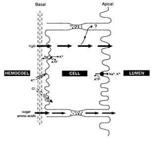 |
Cell types
Rhodnius has two 2 cell types in the Malpighian tubules. Resorption occurs in the proximal region (closest to the gut) leading to the formation of uric acid granules in the Malpighian tubules.
I. Honeycomb border.
Cells with closely packed microvillae (described below). These are the main secretory cells of the tubule and are concentrated in the distal portion of the tubules.
II. Brush border.
Cells with more widely dispersed microvillae (see diagram). Less complex basal invaginations. Mitochondria concentrated basally, suggesting active transport is taking place at the basal surface. concentrated in the proximal portion of the tubules.
Distal (honeycomb): Na and K actively pumped across basal and apical membranes. Water and urate follows across the osmotic gradient. This "primary urine" is isosmotic with the haemolymph.
Proximal (brush): K+ actively pumped into haemolymph, drawing water out and precipitating uric acid granules
Other insects e.g. Carausius have a common cell type along the length of the tubule. Occasionally interspersed specialist cells of unknown function. They may secrete acid mucopolysaccharide.
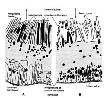 |
Diagrammatic representation of primary Malpighian tubule rells from Rhodnius. (A) A cell from the distal region showing welldeveloped brush border with elongate microvilli, (B) A cell from the proximal region showing the irregular filaments of the brush border and the accumulation of mitochondria in the basal region. Arrows indicate direction of secretion. From Hadley, 1994.) |
Cell morphology
Apical cell surfaces composed of closely packed microvillae, forming an inner brush border. Mitochondria are abundant beneath and sometimes within the microvillae: i.e. an active (energy requiring) area with increased surface area. Suggests membrane associated active transport. The basal cell membrane is deeply invaginated.
Urine production
Uric acid is the most common excretory product because it is non toxic and can be stored.
Urine production is based upon the active generation of osmotic gradients.
Endocrine regulation
In many insects feeding stimulates urine production through a haemolymph-borne hormonal factor: a diuretic hormone.
Best known example is Rhodnius where a blood meal stimulates urine production in the Malpighian tubules and inhibits water uptake in the rectum. In this insect the water content of the meal is immediately reduced to decrease the gut volume. Produce moist faeces.
Antidiuretic hormones typically enhance fluid resorption in the rectum.
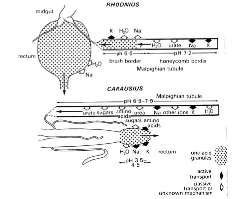
Diagrammatic representation of the movement of inorganic ions and organic molecules into and out from the excretory system of Rhodnius and Carausius (From Chapman).
Rectal absorption
The rectum is a major site of absorption of solutes and water. It is relatively thin walled and lined with a thin, permeable cuticle.
Rectal pad cell of cockroach
- morphology (see diagrams)
- ions are recycled from the intercellular spaces back into the rectal pad cells
- Net flow of water from gut contents -> through cells -> into intercellular spaces -> into haemolymph, against an osmotic gradient.
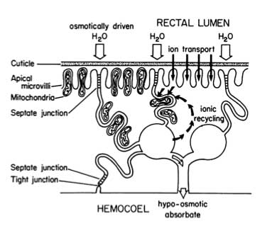 |
Diagrammatic representation of solute and water movements in insect rectal pads during fluid resorption from the rectal lumen. The anatomical organization is shown or the left and the physiological events on the right. Pathways of water movement are indicated by open arrows and solute movements by solid arrows. From Hadley, 1994, adapted Bradley (1985). |
Rectal papillae of fly
- morphology (see diagrams)
- Ions actively transported into intercellular spaces, drawing water from gut lumen into the spaces. On a cellular basis the rectal papilla functions similarly to the rectal pad cells.
- Modified to produce a more circuitous route for the flow of solute from the gut to the haemolymph. Allows further resorption of ions from the solute.
 |
Diagram of the organisation of the rectal papilla of Calliphora. A. Section through part of a papilla showing the direction of movement of water (thin arrows) and solutes (thick arrows). B. The structure of a single epidermal cell in a papilla. (From Chapman, after Berridge, 1970.) |
Cryptonephridial system
- An arrangement of the distal Malpighian tubules in close association with the rectum, surrounded by a perinephric membrane.
- Found in some larval Coleoptera, most larval Lepidoptera, and larval Mycetophilidae + Keroplatidae (mushroom flies, Diptera).
- Diagrams show cell morphology, function, ion and water flow.
- Dual function in Tenebrio: (1) extract water from the hindgut, (2) to absorb water vapour from the air.
- Ions are actively pumped into the Malpighian tubule lumen (perhaps from the haemolymph) and water follows, being drawn from the hindgut lumen, resulting in very dry faeces.
- Flow is reversed in the rectum, causing the hindgut contents to move anteriorly. K+ ions pumped from the haemolymph into the Malpighian tubule lumen through the leptophragma. Water is extracted from the faeces and from the air. An anal valve closes off the rectum when the ambient relative humidity drops.
 |
Left. (A) Anatomical arrangement of "cryptonephridial complex" in tenebrionid beetle larvae (only one of six Malpighian tubules is shown). (B) Schematic transverse section (dashed line in A) of complex showing epithelial and fluid compartments. From Hadley, 1994, adapted from Machin and O'Donnell (1991). Right. A diagram of the cryptonephridial portion of the excretory system of Tenebrio molitor. The entire cryptonephridial complex is surrounded by the perinephric membrane, which serves to osmotically isolate the Malpighian tubules and rectum from the hemolymph. Pathways of ion transport (principally K+ and Cl) are indicated by solid arrows and the direction of passive water diffusion by open arrows. Various routes by which ions might enter the tubules are discussed in the text. (From Hadley 1994, adapted from Bradley, 1985). |
 |
ReferencesHadley, N.F. (1994) Water Relations of Terrestrial Arthropods. QL434.72.H33 Chapman, Ch. 18, Excretion and Salt and Water Regulation
Romoser and Stoffolano “The Science of Entomology” pp 118-123 has a good overview of excretion. |