Insect Hormones
Objectives
- Know the major types of hormones and the types of tissues that produce them.
- Be able to compare the roles of the nervous and hormonal systems in the regulation of insect physiology.
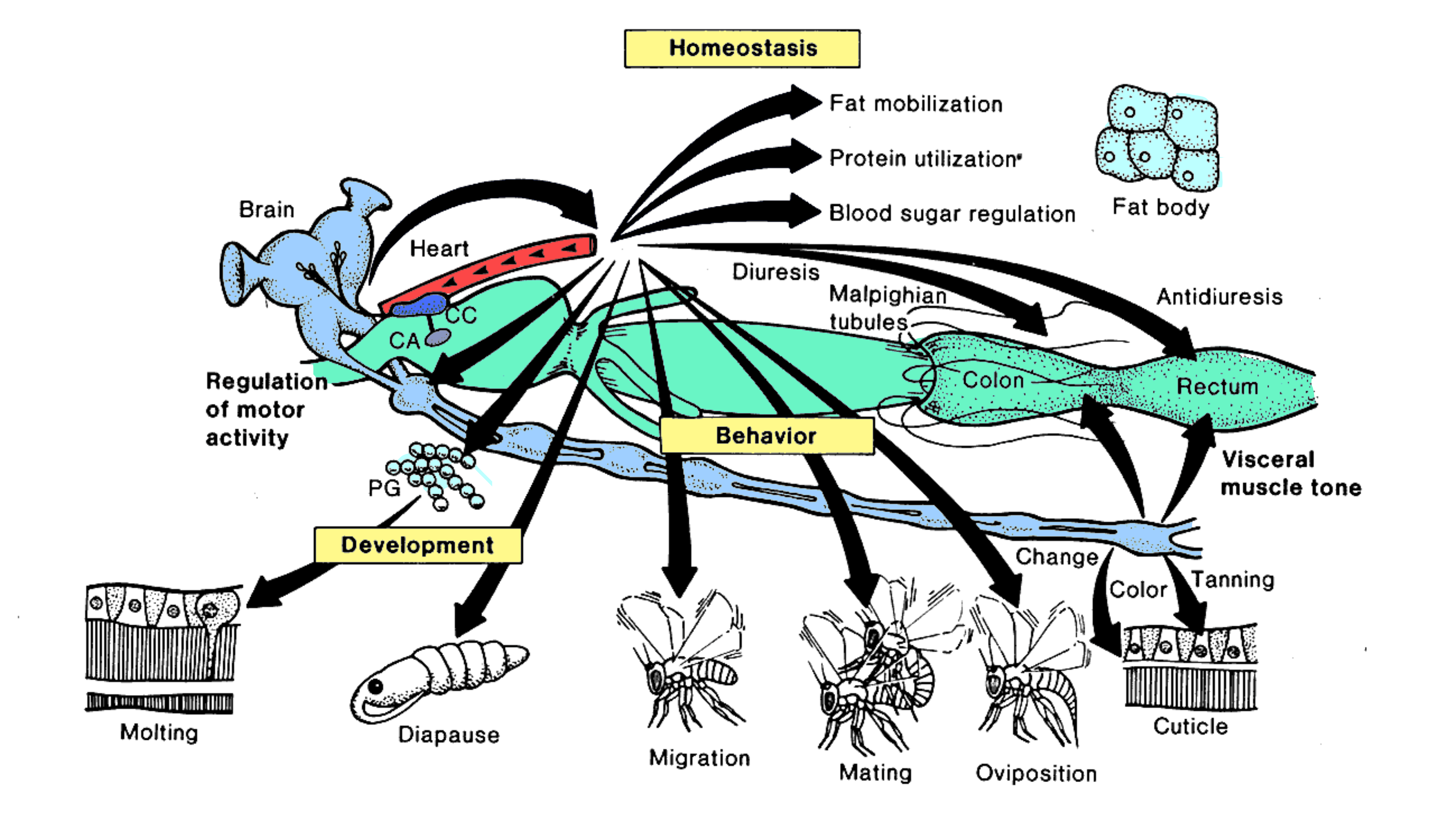
- Be able to present an overview of the types of physiological functions that come under hormonal control.
- you will know the difference between the polyphenism and polymorphism.
Topic Outline
- Part 1: Introduction, Hormone production, hormone types and overview of hormone-modulated processes: moulting and metamorphosis, metabolic activiites, diapause, reproduction
- Part 2: Polyphenisms, Chromatic Adaptations, Phase polyphenism in locusts
Activities
- Minilecture 1: Hormone production systems
- Minilecture 2: Hormones in Moulting and Metamorphosis
- Go to the journal Science and read the following paper by Suzuki and Nijhout
Suzuki, Y. and Nidjout, H.F. 2006. Evolution of a polyphenism by genetic accommodation. Science vol. 311, no. 5761, pp 650-652.
Part 1: Introduction
Hormones play an important regulatory role in insect physiology.
The chemical signal (messenger) enters the circulatory system and is distributed throughout the body. By comparison with the nervous system this is slow and it produces a dispersed rather than localised effect.
It involves a single effector (a gland or group of glands) rather than a highly complex nervous response in which a similar response would have to be hardwired.
Products can be accumulated before distribution (though this doesn't happen in all cases)
Hormones have a coordinating role. Many behavioural and physiological processes can be coordinated by hormonal control. Moulting is an example.
The nervous system is the prime regulator of the hormonal system, thus sensory input—both internal and external—is integrated into the regulation of hormone release.
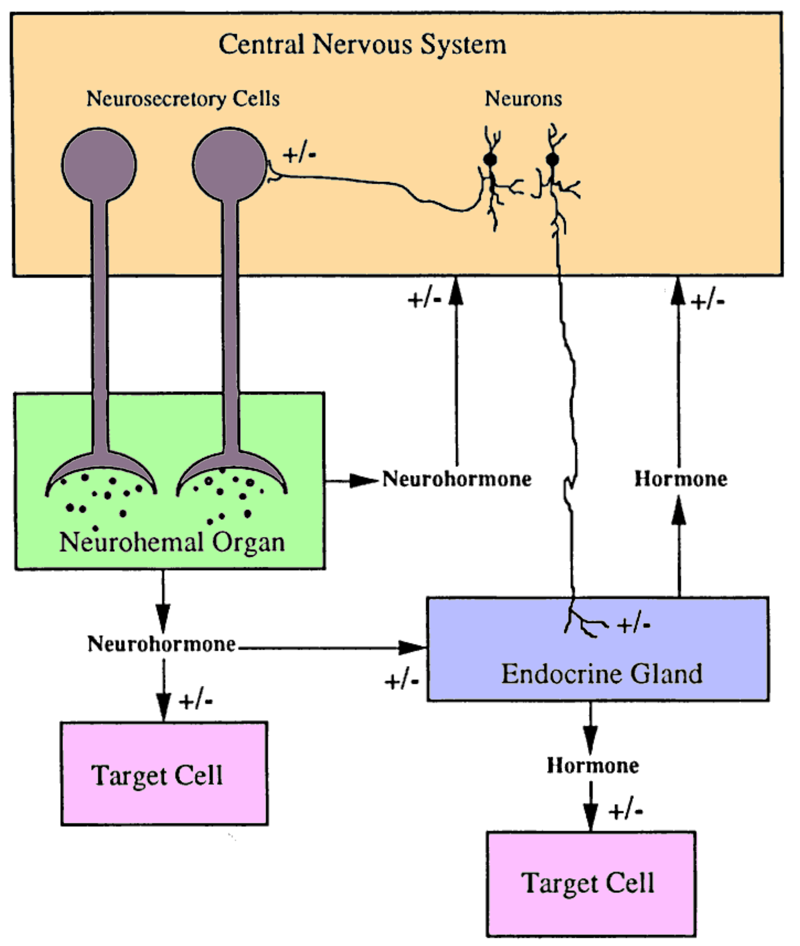 |
A general scheme for pathways of neuroendocrine regulation in insects. The central nervous system is the source of a large variety of neurosecretory hormones. These hormones are released from specialized neurohemal organs and may have an effect directly on a target tissue, or indirectly via the stimulation (+) or inhibition () of the secretory activity of conventional endocrine glands. Neurosecretory hormones can also feed back on the central nervous system and stimulate or inhibit its nervous and neuroendocrine activity. The central nervous system can also stimulate (or inhibit) neurosecretory cells and endocrine glands directly via conventional neurons. From Nijhout, 1994. |
|
Hormone production
Endocrine glands
Two of the main endocrine glands are the prothoracic glands, located in the prothorax (the primary source of ecdysteroids) and the corpora allata (source of juvenile hormones).
Gonads: ovaries and testes
Secretory activity of endocrine glands is controlled via release of neurohormones from the CNS
Neurosecretory cells
Neurosecretory cells are neurons (found primarily in the CNS), that produce polypeptides instead of neurotransmitters.
 The neurons usually have large cell bodies and widely dispersed axons.
Electron-dense granules in cells are usually densely distributed in the cell body and around periphery of nerves and ganglia. Frequently, the nerve terminals are concentrated in special “neurohaemal organs” which are sites of neurohormone release in the peripheral nerves or on the periphery of ganglia.
As integral parts of the nervous system, the neurosecretory cells are under nervous control. These cells form the link between the nervous system and endocrine system.
Techniques:
Ligation |
 |
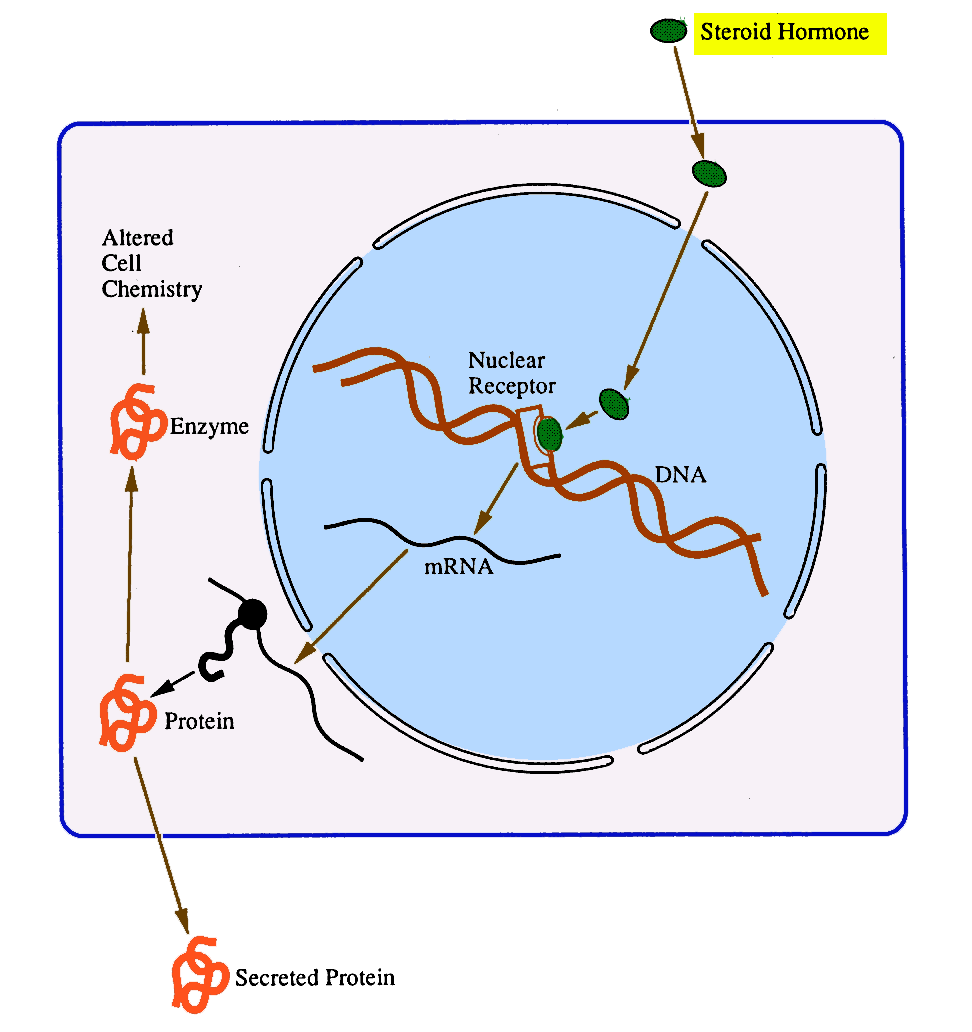
Mechanism of action of a steroid hormone. A steroid hormone passes through the cell membrane, enters the nucleus, and binds to a nuclear receptor protein. The receptor hormone complex then binds to specific regions of the DNA to control gene transcription. From Nijhout, 1994. |
 |
Hormone types
Lipid hormones
Act on genes.
Pass through membranes and bind to receptors within the cell, to DNA for example, and thus can directly regulate transcription.
Ecdysone (a steroid) and juvenile hormone (JH) (a terpenoid) is a lipid hormone
Polytene cells of the Drosophila salivary glands were used to indicate gene transcription in response to ecdysteroids. Ecdysteroid injections cause “puffing” of the polytene chromosomes, indicative of localised transcription. There is a characteristic series of puffs in different locations indicating a cascading series of transcriptional events.
Polypeptides or amines (peptide hormones)
Bind to receptors in cell membrane of the receiving cell
The signal must be carried from the cell membrane where the receptor lies.
Act via a “second messenger” system to activate or depress enzymes or proteins and thus change the physiology of the cell. The second messengers are cAMP or cGMP. Involved in initiating a cascade of protein or enzyme activations that ultimately alter the cell’s physiology.
Hormone-receptor complexes can also act on Ca++ ion concentrations within the cell via a 2nd messenger system
Examples: Eclosion hormone and prothoracicotropic hormone
Peptide hormones usually produced by neurosecretory cells
Brain-retrocerebral complex
Composed of:
|
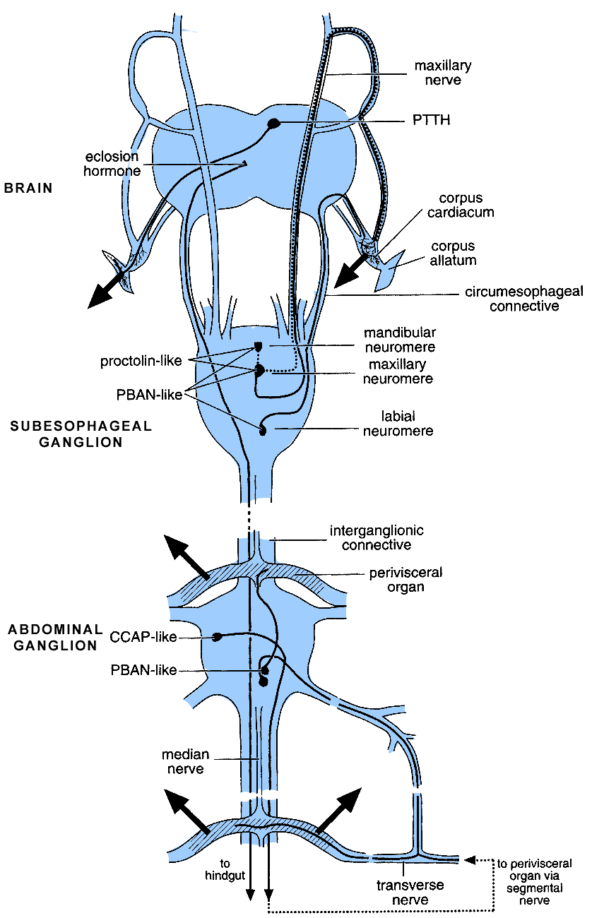 |
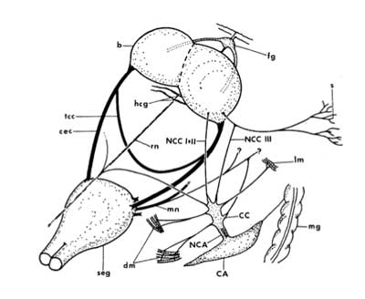
Anatomy of the brain-retrocerebral neuroendocrine complex in a final instar larva of Manduca sexta, showing the anatomical relations of parts and their nervous interconnections. Question marks indicate nerves with unknown destination. (From Nijhout, 1994.) |
b, brain; CA, corpus allatum: CC. corpus cardiacum; cec, circumesophageal connective: dms, dilator muscles of stomodaeum; fg, frontal ganglion: hcg, hypocerebral ganglion; lm, labial muscles; ma, mandibular gland; mn, maxillary nerve; NCA, nervus corporis allati; NCCI+II, nervi corporis cardiaci I and II; NCCIII, nervus corporis cardiaci III; rn, recurrent nerve; s, sensilla; sea, subesophageal ganglion; tcc, tritocerebral commissure.
|
Brain neurosecretory cells |
|
|
Relatively large neurons that produce peptides instead of neurotransmitters. Viewed with the electron microscope the peptide “packets” are large electron-dense vesicles. |
Corpora cardiaca |
|
|
2 functions: |
Corpora allata |
|
|
Glands that are innervated from brain, via the corpora cardiaca |
Prothoracic glands |
|
|
Produce ecdysteroids |
Ventral nerve cord |
|
|
Perivisceral organs: neurohaemal organs for segmental ganglionic neurosecretory cells |
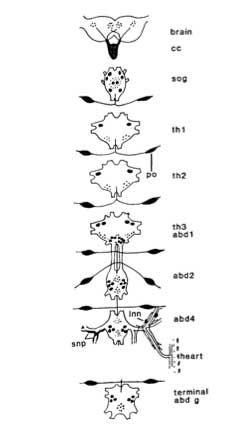 |
Distribution of neurosecretory cells (dots in the ganglia) and anatomical arrangement of neurohemal organs (black swellings on nerves) in a typical insect central nervous system. The neurohemal organs ot the brain are the corpora cardiaca (cc) and those of the abdominal ganglia are the perivisceral organs (po); abd, abdominal ganglia: sog, subesophageal ganglion; th, thoracic ganglia. (From Orchard and Loughton, 1985. Reprinted with kind permission of Pergamon Press Ltd.) From Nijhout, 1994. |
Overview of hormone-modulated processes
1. Moulting and Metamorphosis
Prothoracicotropic hormone |
Released from brain, accumulates in CA, prompts the prothoracic glands to release ecdysone |
|
Ecdysone |
|
|
I. apolysis |
||
Eclosion hormone* |
Released from brain (a neuropeptide) through corpora cardiaca (in Manduca through proctodeal nerve NH area from cells with somata in brain) and targets the CNS to produce eclosion (or ecdysis) behaviour, often timed according to circadian rhythm |
|
Ecdysis triggering hormone* |
Released from peripheral nerve cells associated with the trachea near the spiracles. Trigger the onset of ecdysis behaviour |
|
Bursicon |
Released from neurosecretory cells of thoracic and abdominal neuromeres |
|
Juvenile hormone |
Secreted by endocrine cells of the corpora allata, widespread targets, maintains juvenile stage |
*These hormones will be covered in more depth in the module on Moulting, Part 4: Eclosion and Ecdysis Triggering Hormone.
|
2. Metabolic activities and homeostasis
Adipokinetic hormone |
Polypeptide of 8-10 amino acids |
|
Diuretic hormone |
In Rhodnius, stretch receptors in abdomen monitor abdominal distension following gluttonous meal, leads to increase in urine production. |
|
Proctolin |
Neuropeptide (pentapeptide) both a neurotransmitter and neurohormone |
|
Cardioactive peptides |
released from perivisceral organs |
3. Regulation of Diapause
Diapause |
Arrested development as an adaptation to adverse conditions that has evolved as an adaptation in anticipation of adverse conditions rather than a response to their onset (torpor). Usually involves some stimulus indicative of impending onset. |
|
Embryonic diapause |
|
4. Involvement in Reproduction
Ovarian hormone (ecdysone) |
Released by ovaries and targets fat body where vitellogenin is released for egg production |
|
Juvenile hormone |
|
|
Mating inhibition hormone |
Male accessory glands targets female CNS to make female refractory |
|
Oviposition initiation hormone |
Targets the oviduct to initiate oviposition |
ReferencesChapman. Chapter 21
|


 Minilecture: