Neural Integration
Part 3: wiring the eye and the antenna
At the end of this section you will be able to answer:
- The wiring of the eye is just as important as eye design
- How do you wire the system to tdetect light, colour and motion?
- what is the basic layout of the visual system?
|
The eye: from the photoreceptor to the brain
Review: the ommatidial structure
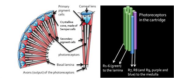
Eight or nine photoreceptors can be within each ommatidium cartridge
The area where the light-detecting microvilli are located is called the rhabdom
They can be open rhabdom or closed rhabdom.
Considering the numerous connections between the sensory input, the motor output, and the internal physiological state: How do we unravel the mechanisms in the ‘black box’ of the insect nervous system?
Photoreceptor arrangementPhotoreceptors 1-6 (labeled R1-6) are often green-sensitive and output to the lamina.
|
 |
The layout of the visual system
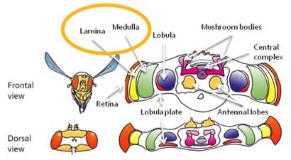
--Retina to lamina to medulla to lobula complex to central brain --
The system is layered within the visual system, but it is also retinotopic, in that the world outside is represented on the retina in a spatial array, with each ommatidial cartridge represented in columns in the lamina, the medulla, and, sometimes, in the lobula complex (see diagram above)
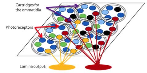
Neural superposition: fly changes in resolution
Since the rhabdom is open, photoreceptors from neighboring cartridges can view the same area of the world. By integrating information across neighboring cartridges, the resolution of the input can be increased.
Photoreceptor inputs and the rest
The lamina inputs (R1-6) are thought to feed into the motion detection pathway.
Meanwhile, the medulla inputs (R7-8) are though to be the major inputs to the color and pattern processing pathway.
More info see: Douglass JK, Strausfeld NJ (1998) Functionally and anatomically segregated visual pathways in the lobula complex of a calliphorid fly. J Comp Neurol 396:84 –104.
The lamina then sends its inputs into the medulla.
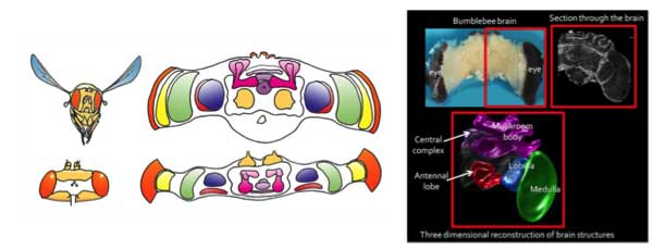
The medulla feeds into the lobula complex, which can be separated into the lobula and lobula plate in many insects (left) or fused (as in bees, right)
Functions: how motion works
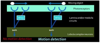
Motion processing involves detecting when objects are moving in the environment.
The way some neurons work is to detect changes in light across neighboring cartridges and integrate it, finally responding when things move in one direction or another (the model is called the Hassenstein-Reichert elementary motion detector).
Borst, A. (2000) Models of motion detection. Nature Neuroscience 3, 1168. doi:10.1038/81435
Motion responses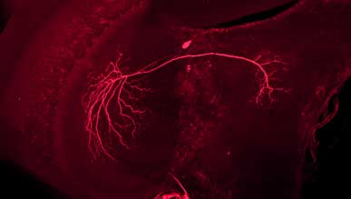
Other neurons can respond to moving bars such as this bumblebee neuron.
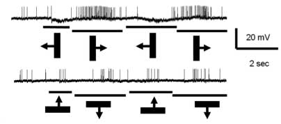
The moving bars are indicated below the membrane potential recording from this cell. You will note that the cell increases its response to the bar moving to the right, but decreases its activity with movement to the left. When the bars move up and down, the cell does not respond.
Different types of cells: wide field versus small field

Cells in the visual system can be wide field (grey and white cells) or small field (black cell) or somewhere in between (white cell).
These different types of visual neurons can perform different roles, such as detecting changes in colour in a small area of the visual field, or detecting motion across the whole visual scene.
Functions: how motion detection works
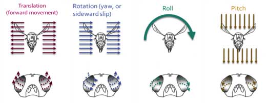
Borst, A. (2000) Models of motion detection. Nature Neuroscience 3, 1168. doi:10.1038/81435
The lobula plate neurons in the fly have been some of the best-studied neurons in any insect.
These neurons are separated into two systems (red arrow below):
The horizontal system, which detects front to back motion, back to front motion
The vertical system, which detects up and down motion
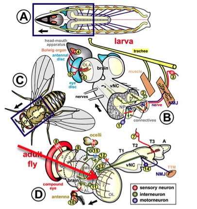
Sánchez-Soriano N, Tear G, Whitington P, Prokop A. Wikipedia Commons
Motion responses in neurons
Still other neurons respond to the movement of small objects, such as for tracking prey:
Neural mechanisms underlying target detection in a dragonfly centrifugal neuron, Read: Bart R. H. Geurten, Karin Nordström, Jordanna D. H. Sprayberry, Douglas M. Bolzon and David C. O’Carroll. The Journal of Experimental Biology 210, 3277-3284. doi:10.1242/jeb.008425
DCMD cell: navigating a swarm
When insects are avoiding a collision or moving through a swarm, like locusts, specific neural circuits allow the insects to avoid collision. Interneurons like the descending contralataral motion detector cell (DCMD) sends inputs to the motor centers to allow the insect to move away from an object.Prof. Claire Rind has some good examples of how collision avoidance circuits can be involved in locust behaviour: http://www.staff.ncl.ac.uk/claire.rind/try1.htm
Dr. Martina Wicklein works on a number of visual systems. Check out her website under ‘current work/preliminary results': http://www.cnl.salk.edu/~martina
Polarized light processing
Polarized light information enters via the photoreceptors, goes through the medulla into a portion of the lobula and projects into the central brain.
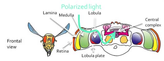 Polarized light information does arrive at the central complex in locusts, where there is a ‘polarization map’ detecting different polarized light directions
Check out this website by Prof. Uwe Homberg, a researcher on how the polarization information is processed in the locust brain:
http://www.uni-marburg.de/fb17/forschung/fobericht/Foberichtneu/homberg
Recommended reading:
Transformation of Polarized Light Information in the Central Complex of the Locust
Stanley Heinze, Sascha Gotthardt, and Uwe Homberg, The Journal of Neuroscience, 29(38):11783-11793; doi:10.1523/JNEUROSCI.1870-09.2009
http://www.jneurosci.org/cgi/content/abstract/29/38/11783
Colour processing
Colour processing is not well understood in insects beyond the photoreceptor level, but evidence has indicated that it is a complex process and can occupy a different pathway from the motion pathway.
- Some cells respond the same way to all the colours the same way, which is called broad-band colour sensitivity.
- Other cells respond to only one or two colours, which is called narrow-band colour sensitivity
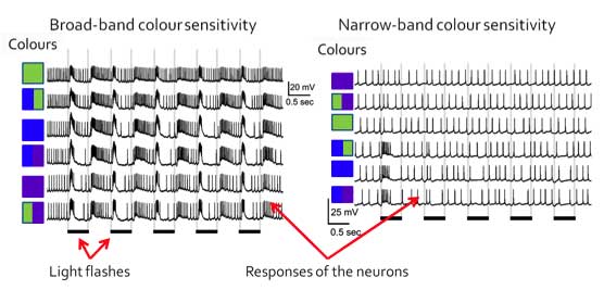
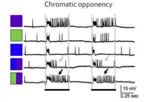
- Some cells respond to the different colours by being excited or inhibited by the different colour combinations, which is called chromatic opponency.
- In this case, the cell was excited by violet, with a number of spikes throughout the light flash.
When the cell was shown violet and blue colour, it produces a burst of spikes in the beginning of the light flash (grey arrows). However, when the cell is shown violet and green, the cell produces a burst of spikes for the duration of the light flash (black arrows). In this case, the blue inhibits the violet response but not the green light, which means that some colours can inhibit or excite responses to other colours.
References:
Hertel H (1980) Chromatic properties of identified interneurons in the optic lobes of the bee. Journal of Comparative Physiology A 137:215-231.
Paulk AC, Phillips-Portillo J, Dacks AM, Fellous JM, Gronenberg W. (2008) The processing of color, motion, and stimulus are anatomically segregated in the bumblebee brain. Journal of Neuroscience 28:6319–6332.
Paulk AC, Dacks AM, Gronenberg W. (2009) Color processing in the medulla of the bumblebee (Apidae: Bombus impatiens). Journal of Comparative Neurology 513: 441-456.
Paulk AC, Dacks AM, Phillips-Portillo J, Fellous JM, Gronenberg W. (2009) Visual processing in the central bee brain. Journal of Neuroscience 29: 9987-9999.
Kien J, Menzel R (1977a) Chromatic properties of interneurons in the optic lobes of the bee-I. Broad-band neurons. Journal of Comparative Physiology. 113:17-34.
Kien J, Menzel R (1977b) Chromatic properties of interneurons in the optic lobes of the bee-II. Narrow band and color opponent neurons. Journal of Comparative Physiology. 113:35-53.
Yang EC, Lin HC, Hung YS (2004) Patterns of chromatic information processing in the lobula of the honeybee, Apis mellifera L. Journal of Insect Physiology 50:913-925.
Functional layout of the visual system
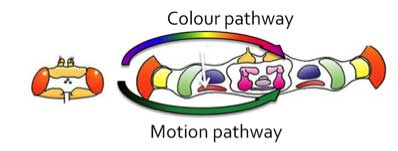
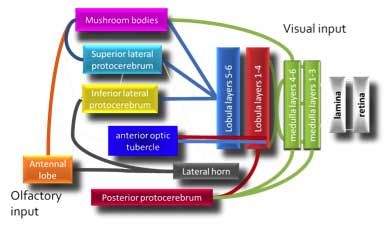
Visual input pathways: where does colour, motion, pattern, and polarized light processing go?
Colour processing can happen in the front of the brain while motion processing may be along the back of the brain.
Smell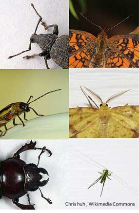
The wiring of the antenna can be essential for olfaction. How do you detect olfactory cues?
What is the basic layout of the olfactory system?
Antennal diversity
There are numerous ways antennae are laid out as is seen in the photos to the right.
The antenna: from the olfactory receptor cells to the brain
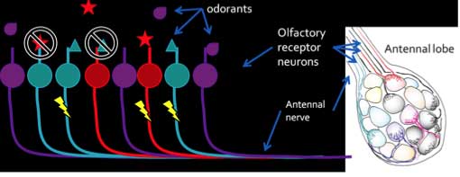
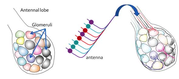
Review: the olfactory receptor cells are located in the antenna, where specific odorants can bind to receptor proteins and induce a spiking response in the olfactory receptors (see Module on Sensing Tastes and Odours: Function) Olfactory receptor cells then form a bundle along the antennal nerve and enter the brain at the antennal lobe.
The ORNs then enter the antennal lobe, where they project to specific structures called glomeruli (spheriodal structures which include synapses and connections between the inputs and the outputs of the antennal lobe). The ORNs of specific odorant receptor types go to the same glomerulus.
Outputs from the antennal lobe
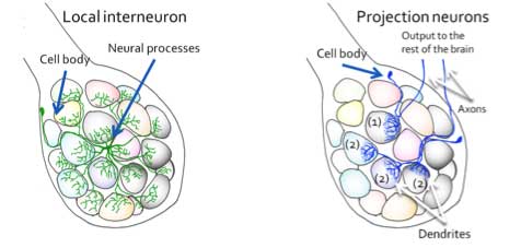
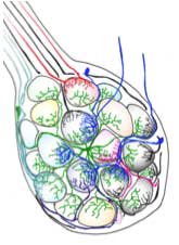
The glomeruli are connected by a number of inhibitory local interneurons (LNs).
The outputs from the antennal lobe are called the projection neurons (PNs), which can receive inputs from single glomeruli (1) or from multiple glomeruli (2).
The PNs project to the lateral horn, in the lateral protocerebrum, and to the mushroom bodies .
Olfactory processing: how does it work?
A lot of debate has occurred in the field as to how olfactory processing may work. The insect models used have been Drosophila melanogaster (fly), Apis mellifera (honeybee), Manduca sexta (tobacco hornworm moth), and the locust (Schistocerca gregaria).
Due to the diversity of the antennal lobe arrangements, there are a very few agreed-upon mechanisms. There is the idea that each glomerulus is an ‘odour’ address, and similar odours are encoded in the same glomerulus, which indicates there could be an odotopic map.
On the other hand, olfactory processing could be a combinatorial type of processing, where each odour has a multiple glomerulus address.
Olfactory and visual processing: where to go from here?
How is all of this information, including mechanosensory information from the thorax and head as well as auditory processing, integrated?
Where does all this information go?
To the protocerebrum and central brain!

Processing in the central brain is where all the information, including mechanosensory, olfactory, auditory, and visual inputs are integrated by the brain.
The central brain areas include the mushroom bodies, the central complex, and the rest of the protocerebrum.
TOPIC REVIEWDo you know…?
|


 Mini-lecture: