Sensing taste and odours
Part 2: Olfaction
|
The olfactory sensilla can be recognised from the multiple pores in their walls. It is hypothesised that these pores let the odour through to the inside of the sensillum. |
Diagram through an olfactory sensillum (smooth-walled peg type). The internal lymph space is fluid-filled. Here the pore opens into a spherical bowl-shaped structure within the wall, referred to as a pore kettle. Epicuticle covers the sensillum and pore opening.
Diagram of a transverse section through a grooved peg showing the channels running from the base of the grooves. [Diagram: B.W. Cribb] |
References
Cribb, B.W. (1997) Antennal sensilla of the female biting midge: Forcipomyia (Lasiohelea) townsvillensis (Taylor) (Diptera: Ceratopogonidae). International Journal of Insect Morphology and Embryology, 24, 405-425.
Shanbhag S.R., Müller, B. & Steinbrecht R.A. (1999) Atlas of olfactory organs of Drosophila melanogaster. Types, external organization, innervation and distribution of olfactory sensilla. International Journal of Insect Morphology and Embryology, 28, 377-397.
Gustation
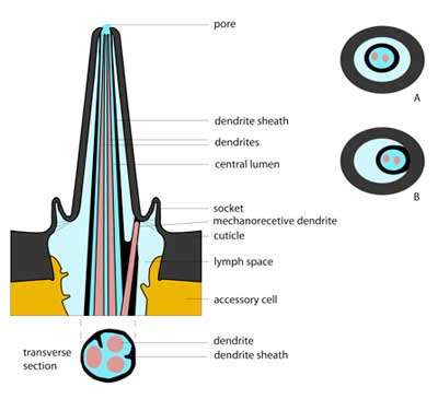
Taste sensilla (gustatory sensilla or contact chemoreceptors) have a single pore at the tip. This pore is bigger than pores in odour-sensitive sensilla and measures about 0.2µm in longest diameter. It can be circular, oval or slit-shaped. A porous plug may fill this hole. |
Styloconic sensillum (peg set on the end of a style) seen on the proboscis of a Helicoverpa armigera moth. Other gustatory sensilla are also present. Image is a scanning electron micrograph. [Image: B.W. Cribb]
|
Within the shaft of the gustatory sensillum is a tube called the dendrite sheath. This houses the dendrite projections from the sensory neurons below. There are different arrangements of the dendrite sheath. It can be a free-standing tube, as seen in grasshoppers (A), or a tube fused to the inside of the sensillum wall over to one side, as in flies (B). |
|
Feeding VideoInsects use some of the body components such as legs and labial/maxillary palps to touch and taste substrates such as food. Where direct contact occurs we are sure to find touch and taste sensilla. The insects shown in this clip are the grasshopper, Valanga irregularis and the Monarch butterfly caterpillar, Danaus plexippus. See if you can see the hairs and spines. Pegs on the very ends of the grasshopper palps are too small to be seen but those further up the segments are visible. Note the long hairs on the antennae of the caterpillar. |


